The same plot in 14 other libraries — JavaScript: D3.js, Apache ECharts, Highcharts, MUI X Charts; Python: Altair, Bokeh, lets-plot, Matplotlib, Plotly, plotnine, Pygal, Seaborn; R: ggplot2; Julia: Makie.jl. Compare all 15 side by side: NMR Spectrum (Nuclear Magnetic Resonance) in Python, R, Julia and JavaScript.
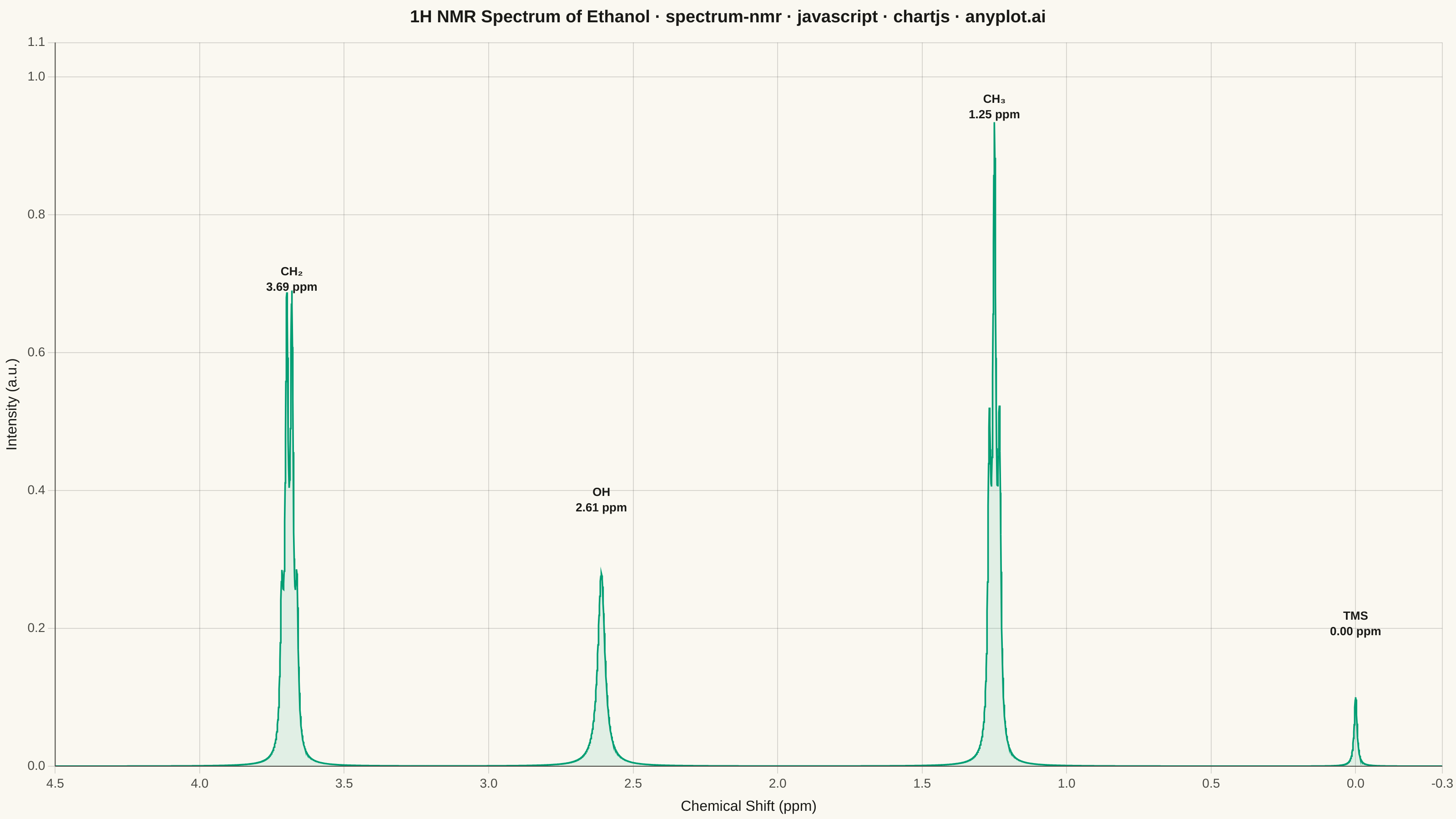
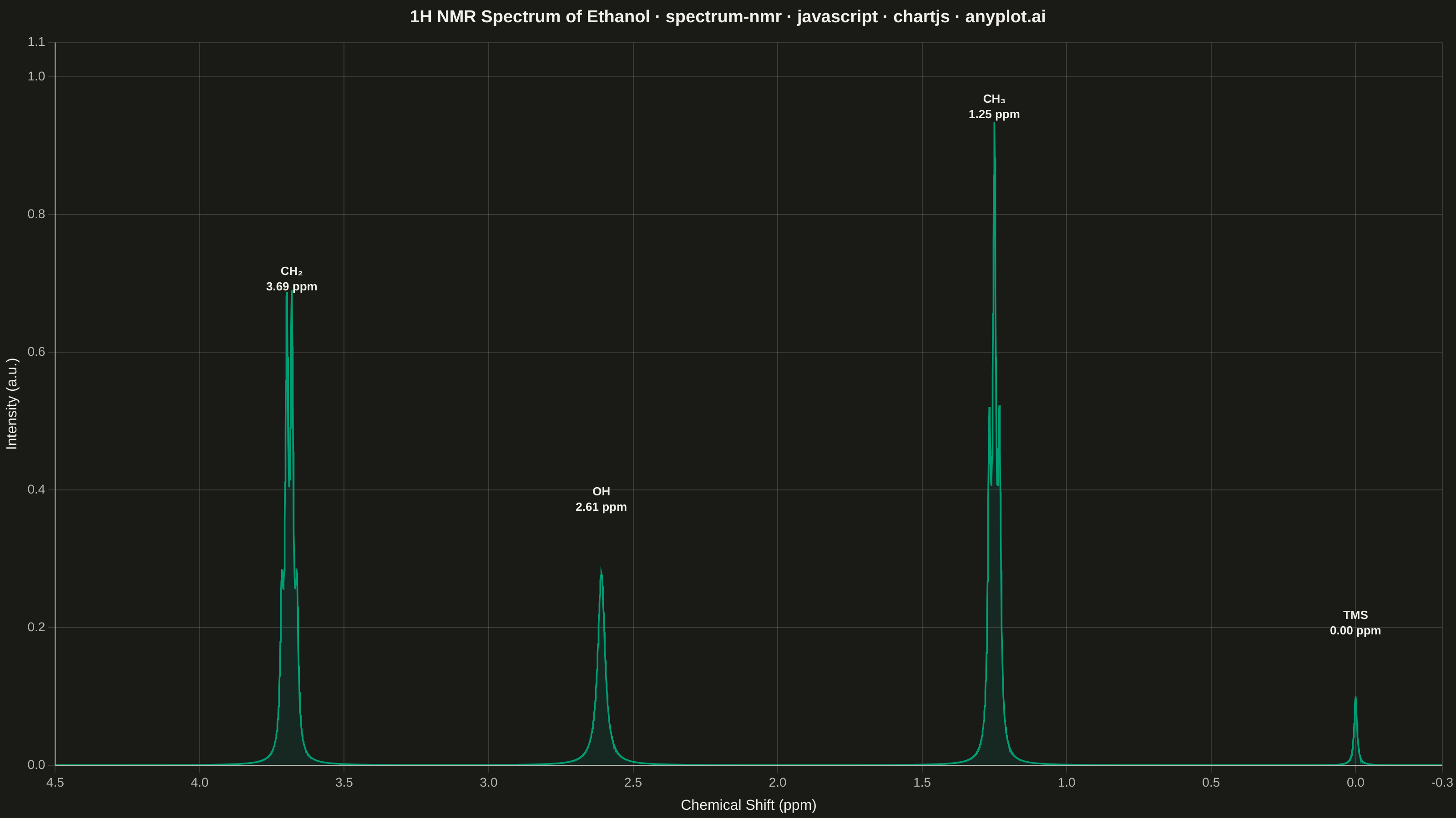
An NMR spectrum plots signal intensity versus chemical shift (in ppm) to reveal the electronic environment of atomic nuclei in a molecule. It is the primary analytical tool in organic chemistry for determining molecular structure, with peak positions indicating functional groups and splitting patterns (multiplets) revealing connectivity between neighboring atoms. The x-axis is conventionally reversed (high ppm on the left) and peaks appear as sharp signals rising from a baseline.
// anyplot.ai
// spectrum-nmr: NMR Spectrum (Nuclear Magnetic Resonance)
// Library: chartjs 4.4.7 | JavaScript 22.22.3
// Quality: 88/100 | Created: 2026-06-03
//# anyplot-orientation: landscape

const t = window.ANYPLOT_TOKENS;

// Lorentzian lineshape: models sharp NMR resonance peaks
const lorentz = (x, c, hw, h) => h / (1 + ((x - c) / hw) ** 2);

// Chemical shift axis: 4.5 to -0.3 ppm, 4000 points
const N = 4000;
const ppmMin = -0.3;
const ppmMax = 4.5;
const ppmStep = (ppmMax - ppmMin) / (N - 1);

// 1H NMR of ethanol (400 MHz): J = 7 Hz = 0.0175 ppm coupling constant
const J = 0.0175;
const hw = 0.006; // Lorentzian half-width (~2.4 Hz at 400 MHz; J/hw ≈ 2.9 gives resolved multiplets)

const peakDefs = [
    // TMS internal standard at 0.00 ppm — singlet, small reference peak
    { c: 0.00,           hw,          h: 0.10, label: "TMS\n0.00 ppm" },
    // CH3 triplet at 1.25 ppm (3H, 1:2:1 intensity pattern)
    { c: 1.25 - J,       hw,          h: 0.42 },
    { c: 1.25,           hw,          h: 0.85, label: "CH₃\n1.25 ppm" },
    { c: 1.25 + J,       hw,          h: 0.42 },
    // OH singlet at 2.61 ppm (1H, broad due to fast proton exchange)
    { c: 2.61,           hw: hw * 2.5, h: 0.28, label: "OH\n2.61 ppm" },
    // CH2 quartet at 3.69 ppm (2H, 1:3:3:1 intensity pattern)
    { c: 3.69 - 1.5 * J, hw,          h: 0.20 },
    { c: 3.69 - 0.5 * J, hw,          h: 0.60, label: "CH₂\n3.69 ppm" },
    { c: 3.69 + 0.5 * J, hw,          h: 0.60 },
    { c: 3.69 + 1.5 * J, hw,          h: 0.20 },
];

// Compute spectrum intensities across the chemical shift axis
const chartData = Array.from({ length: N }, (_, i) => {
    const ppm = ppmMax - ppmStep * i;
    const y = peakDefs.reduce((sum, p) => sum + lorentz(ppm, p.c, p.hw, p.h), 0);
    return { x: ppm, y };
});

// Peaks that carry labels
const labeledPeaks = peakDefs.filter(p => p.label);

// Title with font size scaled to length (baseline 67 chars → 22px)
const titleText =
    "1H NMR Spectrum of Ethanol · spectrum-nmr · javascript · chartjs · anyplot.ai";
const titleFontSize = Math.max(15, Math.round(22 * 67 / titleText.length));

// Fill the full canvas with the page background before Chart.js draws
const bgPlugin = {
    id: "bg",
    beforeDraw({ ctx, width, height }) {
        ctx.save();
        ctx.fillStyle = t.pageBg;
        ctx.fillRect(0, 0, width, height);
        ctx.restore();
    },
};

// Draw only bottom and left axis spines (L-shaped frame, scientific style)
const spinePlugin = {
    id: "spines",
    afterDatasetsDraw({ ctx, chartArea: { top, right, bottom, left } }) {
        ctx.save();
        ctx.strokeStyle = t.inkSoft;
        ctx.lineWidth = 1;
        ctx.beginPath();
        ctx.moveTo(left, top);
        ctx.lineTo(left, bottom);
        ctx.moveTo(left, bottom);
        ctx.lineTo(right, bottom);
        ctx.stroke();
        ctx.restore();
    },
};

// Draw chemical group labels above each key peak
const labelPlugin = {
    id: "peakLabels",
    afterDatasetsDraw({ ctx, scales: { x: xs, y: ys } }) {
        ctx.save();
        ctx.textAlign = "center";
        ctx.fillStyle = t.ink;
        ctx.font = "600 13px sans-serif";
        labeledPeaks.forEach(({ c, h, label }) => {
            const px = xs.getPixelForValue(c);
            const lines = label.split("\n");
            lines.forEach((line, i) => {
                const py = ys.getPixelForValue(h + 0.09) - (lines.length - 1 - i) * 17;
                ctx.fillText(line, px, py);
            });
        });
        ctx.restore();
    },
};

// Mount
const canvas = document.createElement("canvas");
document.getElementById("container").appendChild(canvas);

new Chart(canvas, {
    type: "scatter",
    data: {
        datasets: [{
            data: chartData,
            showLine: true,
            borderColor: t.palette[0],
            backgroundColor: t.palette[0] + "1a",
            borderWidth: 1.8,
            pointRadius: 0,
            fill: "origin",
            tension: 0,
        }],
    },
    options: {
        responsive: true,
        maintainAspectRatio: false,
        animation: false,
        plugins: {
            title: {
                display: true,
                text: titleText,
                color: t.ink,
                font: { size: titleFontSize, weight: "600" },
                padding: { top: 8, bottom: 16 },
            },
            legend: { display: false },
            tooltip: { enabled: false },
        },
        scales: {
            x: {
                type: "linear",
                reverse: true,
                min: ppmMin,
                max: ppmMax,
                border: { display: false },
                ticks: {
                    color: t.inkSoft,
                    font: { size: 14 },
                    stepSize: 0.5,
                    callback: (v) => v.toFixed(1),
                },
                grid: { color: t.grid },
                title: {
                    display: true,
                    text: "Chemical Shift (ppm)",
                    color: t.ink,
                    font: { size: 16 },
                },
            },
            y: {
                min: 0,
                max: 1.05,
                border: { display: false },
                ticks: {
                    color: t.inkSoft,
                    font: { size: 14 },
                    callback: (v) => v.toFixed(1),
                },
                grid: { color: t.grid },
                title: {
                    display: true,
                    text: "Intensity (a.u.)",
                    color: t.ink,
                    font: { size: 16 },
                },
            },
        },
    },
    plugins: [bgPlugin, spinePlugin, labelPlugin],
});
Runnable source as JSON, for any HTTP client: https://api.anyplot.ai/specs/spectrum-nmr/chartjs/code. Any spec id and library id listed in llms-full.txt fit the same URL shape; every URL below is complete and callable.
{
  "spec_id": "spectrum-nmr",
  "language": "javascript",
  "library": "chartjs",
  "page": "https://anyplot.ai/spectrum-nmr/javascript/chartjs",
  "hub": "https://anyplot.ai/spectrum-nmr",
  "code_json": "https://api.anyplot.ai/specs/spectrum-nmr/chartjs/code",
  "spec_json": "https://api.anyplot.ai/specs/spectrum-nmr",
  "render_light_png": "https://storage.googleapis.com/anyplot-images/plots/spectrum-nmr/javascript/chartjs/plot-light.png",
  "render_dark_png": "https://storage.googleapis.com/anyplot-images/plots/spectrum-nmr/javascript/chartjs/plot-dark.png",
  "interactive_light_html": "https://storage.googleapis.com/anyplot-images/plots/spectrum-nmr/javascript/chartjs/plot-light.html",
  "interactive_dark_html": "https://storage.googleapis.com/anyplot-images/plots/spectrum-nmr/javascript/chartjs/plot-dark.html",
  "quality_score": 88.0,
  "license": "MIT",
  "guide": "https://anyplot.ai/llms.txt"
}
Part of NMR Spectrum (Nuclear Magnetic Resonance) on anyplot.ai.